
Our Group’s research mainly focuses on fundamental understanding of nano-bio interactions in vitro and in vivo and further applying these understandings to advance disease diagnosis and therapy.
(I) Nanoparticle transport and interactions in-vivo
(II) Biomedical applications of renal clearable metal nanoparticles
(III) Nanochemistry and photophysics for luminescent metal nanoparticles
(I) Nano-particle transport and interactions in vivo
a. Previously unknown proximal tubular elimination mechanism

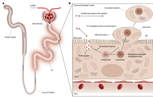
Proximal tubular cells form balloon-like structures in their luminal membranes that contained not only gold nanoparticles but also lysosomes, mitochondria, endoplasmic reticulum and other organelles typically confined to a cell’s interior. The extruded contents were then pinched off into a vesicle that floated off into the extracellular space. This is a previously unknown “housekeeping” process in kidney cells that ejects unwanted content, resulting in cells that rejuvenate themselves and remain functioning and healthy.
Recent Publications:
Yingyu Huang, Mengxiao Yu, Jie Zheng. Nature Nanotechnology 2023. DOI: 10.1038/s41565-023-01366-7
b. Glomerular filtration of few-atom gold nanoclusters: Size effect
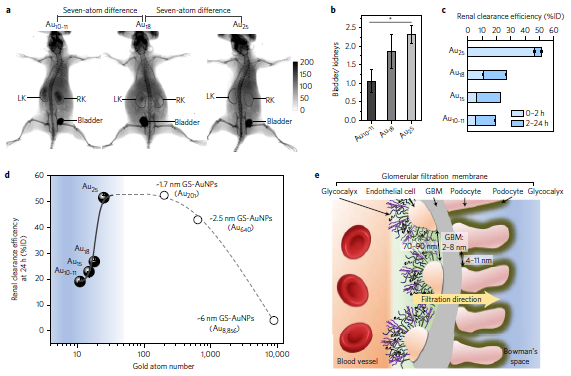
Glomerular filtration barrier behaves as an atomically precise ‘bandpass’ filter to significantly slow down renal clearance of few-atom gold nanoclusters (AuNCs) with the same surface ligands but different sizes (Au18, Au15 and Au10-11).Compared to Au25, just few-atom decreases in size result in four-to-nine fold reductions in renal clearance efficiency in the early elimination stage, because the smaller AuNCs are more readily trapped by the glomerular glycocalyx than larger ones.
Recent Publications:
Bujie Du, Xingya Jiang, Anindita Das, Qinhan Zhou, Mengxiao Yu, Rongchao Jin, Jie Zheng, Nature Nanotechnology, 2017 DOI: 10.1038/nnano.2017.170
b. In vivo transport of renal clearable metal nanoparticles in the kidneys: Margination effect
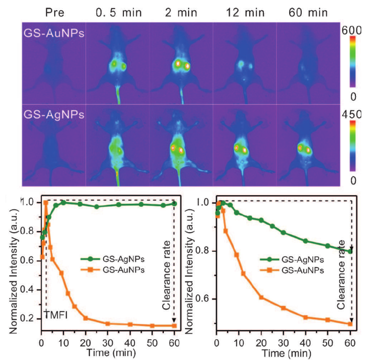
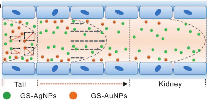
Lower-density NPs are more easily distributed in the body andhave shorter retention times in highly permeable organs thanhigher-density NPs. The density-dependent in vivo behavior ofmetal NPs likely results from their distinct margination in laminarblood flow, which opens up a new path for precise control ofnanomedicines in vivo.
Recent Publications:
Tang, S., Peng, C., Xu, J., Du, B., Wang, Q., Vinluan, R. D., Yu, M., Kim, M.J., Zheng, J. Tailoring Renal Clearance and Tumor Targeting of Ultrasmall Metal Nanoparticles with Particle Density. Angew. Chem. Int. Ed., 2016, 55, 16039-16043.
c. Tumor microenvironment: Vasculature and Acidity Effects
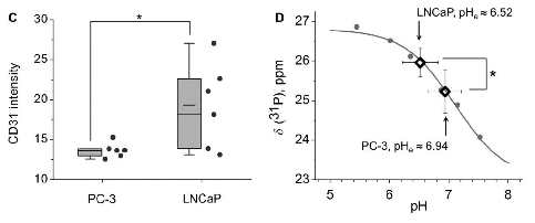
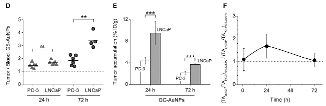
Increased tumor acidity indeed enhanced the uptake of AuNPs with acidity targeting, but only for a limited period oftime. By making use of simple surface chemistry, these two effects can be synchronized in time for high tumortargeting, opening new possibilities to further improve the targeting efficiencies of nanomedicines.
Recent Publications:
Yu, M., Zhou, C., Liu, L., Zhang, S., Sun, S., Hankins, J.D., Sun, X. and Zheng, J., 2017. Interactions of Renal-Clearable Gold Nanoparticles with Tumor Microenvironments: Vasculature and Acidity Effects. Angewandte Chemie, 129(15), pp.4378-4383.
(II) Biomedical applications of renal clearable metal nanoparticles
To investigate whether renal clearable metal NPs can target and image tumors with poor permeability, we used an orthotopic murine glioma model to investigate the passive targeting of 3 nm renal clearable gold nanoparticles (AuNPs) and found out that 3-nm AuNPs were able to target intracranial tumor tissues with higher efficiency (2.3× relative to surrounding non-tumor normal brain tissues) and greater specificity (3.0×) than did the larger AuNPs
Recent Publications:
Peng, C., Gao, X., Xu, J., Du, B., Ning, X., Tang, S., Bachoo, R.M., Yu, M., Ge, W.P. and Zheng, J., 2017. Targeting orthotopic gliomas with renal-clearable luminescent gold nanoparticles. Nano Research, 4(10), pp.1366-1376.
b. Noninvasive fluorescence kidney functional imaging
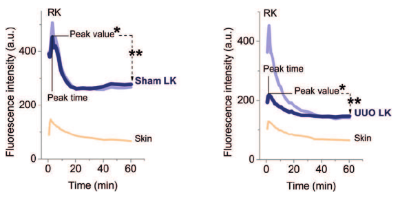
To address the long-standing challenge in noninvasive fluorescence kidney imaging, we are developing renal clearable metal nanoparticles for noninvasive imaging of kidney function and noninvasive staging of kidney dysfunction.
Recent Publications:
(1) M. X. Yu, J. C. Zhou, B. J. Du, X. H. Ning, C. Authement, L. Gandee, P. Kapur, J. T. Hsieh, J. Zheng. “Noninvasive Staging of Kidney Dysfunction Enabled by Renal Clearable Luminescent Gold Nanoparticles”,Angew. Chem. Int. Ed., 55, 2787-2791 (2016) (Highlighted as VIP).
(2) M. X. Yu, J. B. Liu, X. H. Ning, J. Zheng*. “High-contrast Noninvasive Imaging of Kidney Clearance Kinetics Enabled by Renal Clearable Nanofluorophores”. Angew. Chem. Int. Ed., 2015, 54, 15434-15438
(3) M. X. Yu, J. Zheng*. “Clearance Pathways and Tumor Targeting of Imaging Nanoparticles”. ACS Nano, 2015, 9, 6655–6674.
c. Multimodality in vivo Imaging
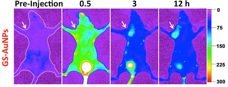
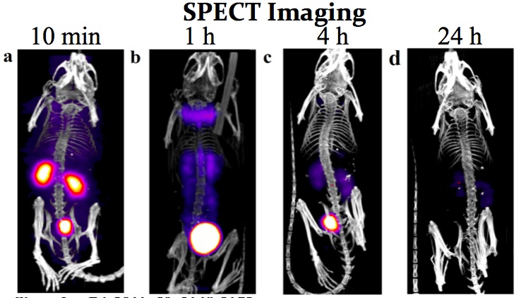
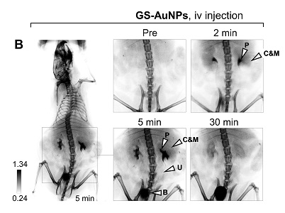
To minimize nanotoxicity, we have developed a class of luminescent metal nanoparticles that can be cleared from the body through urinary system but still serve as a new generation of contrast agents for early disease diagnosis.
Recent Publications:
(1) J. Xu, M. Yu, P. Carter A, E. Hernandez, A. Dang, P. Kapur, J. T. Hsieh, J. Zheng. “In Vivo X-ray Imaging of Transport of Renal Clearable Gold Nanoparticles in the Kidneys”, Angew. Chem. Int. Ed., 2017,DOI: 10.1002/anie.201707819 (online) (Highlighted as VIP)
(2) J. Liu, M. Yu, C. Zhou, S. Yang, X. Ning, and J. Zheng, “Passive Tumor Targeting of Renal Clearable Luminescent Gold Nanoparticles: Long Tumor Retention and Fast Normal Tissue Clearance”, J. Am. Chem. Soc., 2013, 135(13), 4978-4981.
(3) C. Zhou, G. Hao, T. Patrick, J. Liu, M. Yu, S. Sun, O. Oz, X. Sun, and J. Zheng, “Near Infrared Emitting Radioactive Gold Nanoparticles with Molecular Pharmacokinetics”, Angew. Chem. Int. Ed., 2012, 51(40), 10118-10122.
(4) C. Zhou, M. Long, Y. Qin, X. Sun and J. Zheng, “Luminescent Gold Nanoparticles with Efficient Renal Clearance”, Angew. Chem. Int. Ed., 2011, 50(14), 3168-3172.
d. Targeting acidic tumor microenvironment and cancer receptor
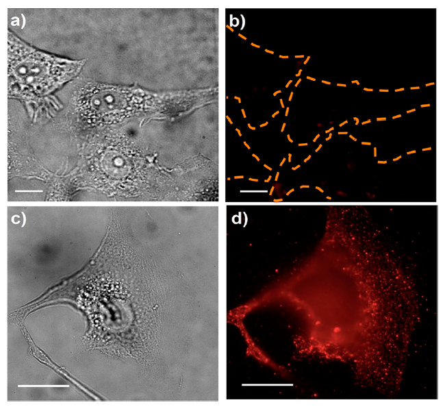
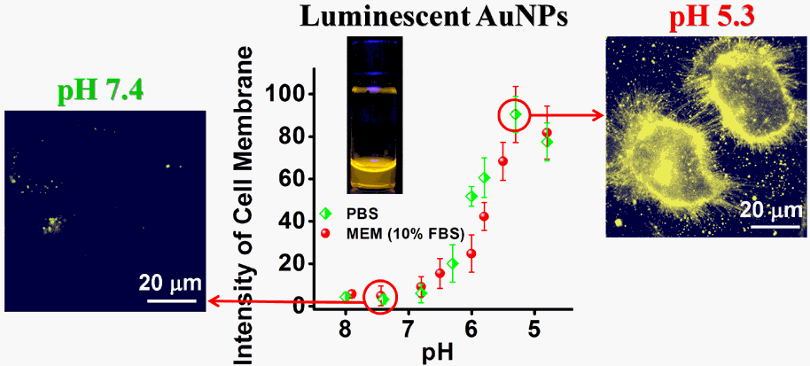
By taking advantage of robust single-particle fluorescence and giant Raman enhancements of unique polycrystalline silver NPs (AgNPs), we found specific c-RGD peptide−αvβ3 integrin interactions not only induced endosome formation more rapidly, enhanced constrained diffusion, but also minimized nonspecific chemical interactions between the NPs and intracellular biomolecules than passive PEGylation chemistry.
Recent Publications:(1) S. Sun, X. Ning, G. Zhang, Y.C. Wang, C. Peng, J. Zheng. “Dimerization of Organic Dyes on Luminescent Gold Nanoparticles for Ratiometric pH Sensing”, Angew. Chem. Int. Ed., 2016, 55, 2421 –2424.(2) S. Sun, C. Zhou, S. Chen, J.B. Liu, J. Yu, J. Chilek, L. Zhao, M.X. Yu, R. Vinluan, B. Huang, and J. Zheng, “Surface-Chemistry Effect on Cellular Response of Luminescent Plasmonic Silver Nanoparticles”, Bioconjugate Chemistry, 2014, DOI: 10.1021/bc500008a.
(3) M. Yu,† C. Zhou,† J. Liu, J. D. Hankins, J. Zheng, “Luminescent Gold Nanoparticles with pH De-pendent Membrane Adsorption”, J. Am. Chem. Soc., 133, 11014-11017 (2011).
(III) Nanochemistry and photophysics for luminescent metal nanoparticles
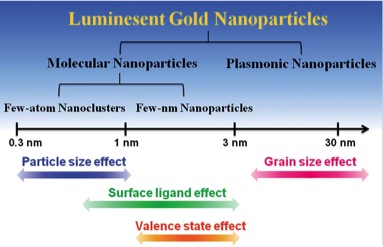
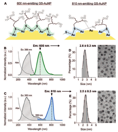
After a decade’s effort, we have developed three synthetic strategies to create luminescent metal nanoparticles with tunable size and surface chemistries. We found out that the emission of luminescent gold nanoparticles is size-independent but related with the conformation of surface ligands. Using structure-fluorescence relationships we discovered, we are now designing a new generation of luminescent metal nanoparticles that can address the challenges in biomedical and energy research.