Prof. Katherine Brown pursues innovation in the monitoring of cancer treatments with super-resolution ultrasound imaging. Signal processing, image processing, and neural networks are the methods used in localizing an ultrasound contrast agent at resolutions up to 10 times smaller than the diffraction limit.
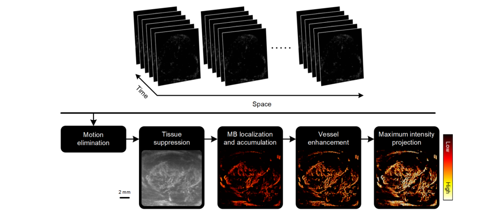
Super-resolution ultrasound images of a microbubble contrast agent flowing in a horizontal channel using a variety of pulsing strategies (vertical axis) and accumulated over 2400 (left column) or 7200 frames (right column). A mixed pulsing strategy of B-mode ultrasound imaging combined with nonlinear contrast imaging (B800 & C) produces a saturated channel at 2400 frames (8 seconds) similar to a single B-mode ultrasound imaging strategy (B-mode) at 7200 frames (24 seconds), representing a 3x speedup.Diagram of the image processing strategy for generating super-resolution ultrasound (SR-US) images. Sequences of ultrasound images (N = 3000 frames) undergo motion elimination then spatiotemporal filtering using a singular value decomposition (SVD) algorithm before microbubble (MB) localization and enumeration to form the final SR-US map. A maximum intensity projection (MIP) is then applied to all spatial SR-US images (N = 6).