
We are grateful to collaborate with incredible engineers, clinicians, and scientists! View them here:
Current & Ongoing Work
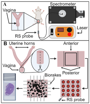
Biomechanical and Compositional Changes in the Murine Uterus with Age
The uterus is a hollow, fibromuscular organ involved in physiologic processes such as menstruation and pregnancy. The content and organization of extracellular matrix constituents such as fibrillar collagen dictates passive (non-contractile) biomechanical tissue function; however, how extracellular matrix composition and biomechanical function change with age in the uterus remains unknown. This study utilizes Raman spectroscopy coupled with biaxial inflation testing to investigate changes in the murine uterus with age (2-3 months, 4-6 months, 10-12 months, and 20-24 months). Additionally, the presented workflow couples biaxial inflation testing and Raman spectroscopy, representing a critical first step to correlating biomechanics and optical signatures in the aging uterus with the potential for clinical translation. Further, this study may provide critical compositional and structure-function information regarding age-related uterine disorders. This project is in collaboration with the Pence Lab at UTSW.
Read the publication in Annals of Biomedical Engineering here!

This work is funded, in part, by NSF CMMI 2053851 (De Vita, Abramowitch, Miller, Myers), UT Rising STARs Award (Pence), AHA Second Century Early Faculty Independence Award 23SCEFIA1156385 (Pence) and AAOGF/Burroughs Wellcome Career Development Award (Florian-Rodriguez).
Elastic Fiber Regeneration Potential During Postpartum Healing
While the etiology of pelvic floor disorders is unknown, vaginal delivery and injuries during childbirth are leading risk factors. Additionally, women with first pregnancies after age 35 have a 350% greater risk of obstetric injury compared to 25-year-olds, yet, nine times more women are delaying childbearing compared to 4 decades ago. The etiology of increased trauma risk is unknown. However, with advancing age, pelvic floor tissue’s reduced elasticity and regenerative capability may contribute. Prior work suggests that regeneration and remodeling of elastic fibers during pregnancy and postpartum healing is critical to pelvic floor disorder prevention. Thus, decreased elastic fiber functionality and regenerative potential with advanced maternal age may contribute to an increased risk of injury during childbirth and, consequently, pelvic floor disorder development. There is a need to rigorously quantify relationships between elastic fibers, contractility, and mechanical properties during postpartum healing as a function of maternal age. Such information will improve our basic science understanding of the regenerative potential of the vagina to produce and remodel elastic fibers, as well as the role of elastic fibers in dynamic remodeling during pregnancy and postpartum healing. This work is being performed in collaboration with the Bersi Lab at WashU.

This work is funded by NSF Award #: 2326797
A Noninvasive Tool to Characterize Evolving Structure-Function Relationships in a Murine model of Pelvic Organ Prolapse
Methods to decipher POP’s pathophysiology commonly involve excising tissue; however, biopsies are invasive and can compromise the biomechanical integrity of the vagina. Therefore, there is a significant need for a noninvasive tool to provide fundamental advances in the scientific knowledge of evolving structure-function vaginal relationships. This study couples Raman spectroscopy and planar biaxial biomechanical testing to compare the well-established fibulin-5 global knockout mouse of POP to the wild type C57BL6 mouse. If successful, this project will yield a new noninvasive assessment technique for prolapse and fundamental knowledge of composition specific tissue remodeling during POP. This project is in collaboration with the Pence Lab and Florian Lab at UTSW.
Biaxial Mechanical Properties and Composition of the Human Vagina in Patients with and without Pelvic Organ Prolapse
Pelvic organ prolapse (POP) is the descent of the female pelvic organs, such as the uterus, bladder, and rectum, into the vagina. While the etiology of POP is not fully understood, loss of mechanical integrity of the vagina, uterosacral ligaments, and other muscular and connective tissues may contribute to POP development and progression. 2.4% of premenopausal and more postmenopausal women require surgical intervention to restore normal anatomy and relieve symptoms such as voiding dysfunctions. The structural and compositional changes that dictate the loss of mechanical integrity are not fully understood, and research on vaginal tissue is limited. In addition, the relationship between most mechanical properties, biochemical composition, and demographics in premenopausal vaginal tissue remains unknown. Therefore, we aim to quantify biaxial mechanical behavior and biochemical content in vaginal tissue from premenopausal and postmenopausal women with and without prolapse. This project is in collaboration with Dr. Leise R. Knoepp, Lyndsey R. Buckner, Dr. Laurephile Desrosiers, and Dr. Maria Florian-Rodriguez.

Figure 1. (A) Samples were collected from apical anterior vaginal wall; (B) planar biaxial testing device cyclically stretched the sample mounted by fishhooks; (C) Vaginal tissue has nonlinear mechanical response (black dashed line: strain-stress relationship) to external load, and a bilinear function (blue line) fitted the curve to quantify moduli (slopes of the region) and extensibility (strains at the transition point and peak).
This work is funded by Ochsner Translational Medicine Research Initiative and UT Dallas CoBRA.
Past Work
Biaxial Murine Vagina Remodeling with Reproductive Aging
Higher reproductive age is associated with an increased risk of gestational diabetes, pre-eclampsia, and severe vaginal tearing during delivery. Further, menopause is associated with vaginal stiffening. However, the mechanical properties of the vagina during reproductive aging before the onset of menopause are unknown. Therefore, the first objective of this study was to quantify the biaxial mechanical properties of the nulliparous murine vagina with reproductive aging. Menopause is further associated with decreased elastic fiber content, which may contribute to vaginal stiffening. Hence, our second objective was to determine the effect of elastic fiber disruption on the biaxial vaginal mechanical properties. To accomplish this, vaginal samples from CD-1 mice aged 2–14 months underwent extension-inflation testing protocols (n = 64 total; n = 16/age group). Then, half of the samples were randomly allocated to undergo elastic fiber fragmentation via elastase digestion (n = 32 total; 8/age group) to evaluate the role of elastic fibers.

This work is funded by NSF Award #: 1947770.
Role of Fibulin-5 Insufficiency and Prolapse Progression on Murine Vaginal Biomechanical Function
Herein, we are extending our work in POP to better understand the interaction between elastic fibers and smooth muscle contractility in the vagina. To accomplish this, we are leveraging the Fibulin-5 deficient mouse model, which readily develops prolapse similar to humans and non-human primates. To address this gap, we are extending our past extension-inflation protocol to assess maximum contractility and the passive biaxial mechanical. These experiments are critical to understanding the potential structural and mechanical processes of POP progression atin to design and improve biomaterial interventions that better recapitulate native function, as well as to develop pharmaceutical interventions.

This work is funded by the NSF Early Faculty CAREER Development Award (BMMB-1751050)
Uterosacral ligaments (USLs) provide structural support to the female pelvic floor, and a loss of USL structural integrity or biomechanical function may induce pelvic organ prolapse (POP). Alterations in extracellular matrix composition and organization dictate USL mechanical function. However, changes in USL microstructure and corresponding mechanical properties still need to be fully understood, and how microstructure and mechanics change with the onset and progression of POP needs to be understood. This is due, in part, to the fact that USL properties are primarily characterized along a single direction (uniaxial test). In contrast, the USL is loaded in multiple directions simultaneously within the body. Biaxial testing permits the acquisition of biomechanical data from two axes simultaneously and thus simulates a more physiologic assessment than traditional uniaxial testing. Therefore, this study aimed to quantify the biaxial biomechanical properties and histological composition of the USL in post-menopausal women with and without POP at various stages. We obtained uterosacral ligament samples from postmenopausal women with either POP or non-POP status following transvaginal hysterectomies with IRB approval in collaboration with Ochsner Clinical School. This information will be a critical component in improving existing finite element models to determine prolapse’s structural and mechanical mechanisms and improve existing clinical treatments.

This work is funded by Ochsner Translational Medicine Research Initiative.