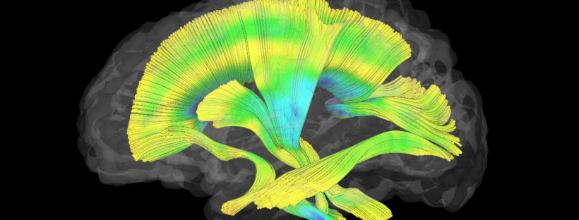
CHAPEL HILL, NC – For the first time, UNC School of Medicine researchers have used MRIs to show that babies with the neurodevelopmental condition fragile X syndrome had less-developed white matter compared to infants that did not develop the condition. Imaging various sections of white matter from different angles can help researchers focus on the underlying brain circuitry important for proper neuron communication.
The study, published in JAMA Psychiatry, shows that there are brain differences related to the neurodevelopmental disorder established well before a diagnosis is typically made at age three or later.